
Endoscopic Spine Surgery
A state-of-the-art surgical approach performed by Dr. Achal Gupta at Neuron Brain & Spine Centre, Lucknow. Utilizing 4K high-definition fluid-irrigated endoscopy through a 7–10 mm micro-incision, targeted neural decompression is achieved with zero muscle resection.
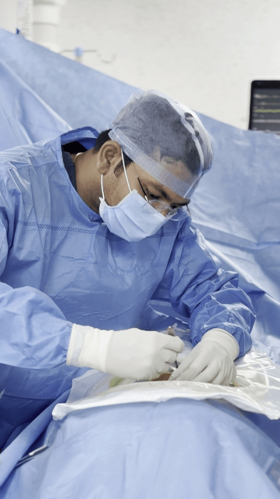
Continuous fluid irrigation keeps the surgical corridor clear while gently separating compressed nerve roots.
Interactive Surgical Corridor Visualizer
Interactive cross-section comparing keyhole muscle dilation with open tissue resection
Endoscopic Spine Surgery vs. Traditional Open Surgery
See the simple difference: How Dr. Achal Gupta treats slipped discs and sciatica through a tiny 7mm keyhole instead of big open cuts
Endoscopic Spine Surgery
Tiny 7mm Band-Aid size puncture • Zero cutting of back muscles • Walk comfortably the same day.
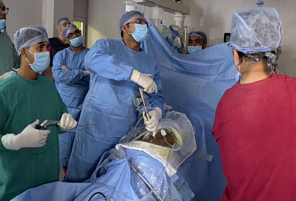
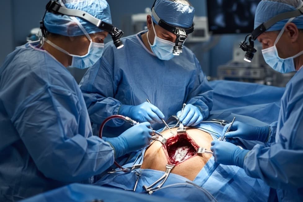
Traditional Open Spine Surgery
Large 70–100mm open incision • Back muscles stripped and cut away • Multi-day hospital bed rest.
Diagnostic Disc & Sciatica Nerve Visualizer
Inspect how disc herniation pinches spinal nerves and maps down the lower limb
Visualizing Spinal Disc Conditions
Interactive axial disc cross-sections illustrating mechanical nerve root compression
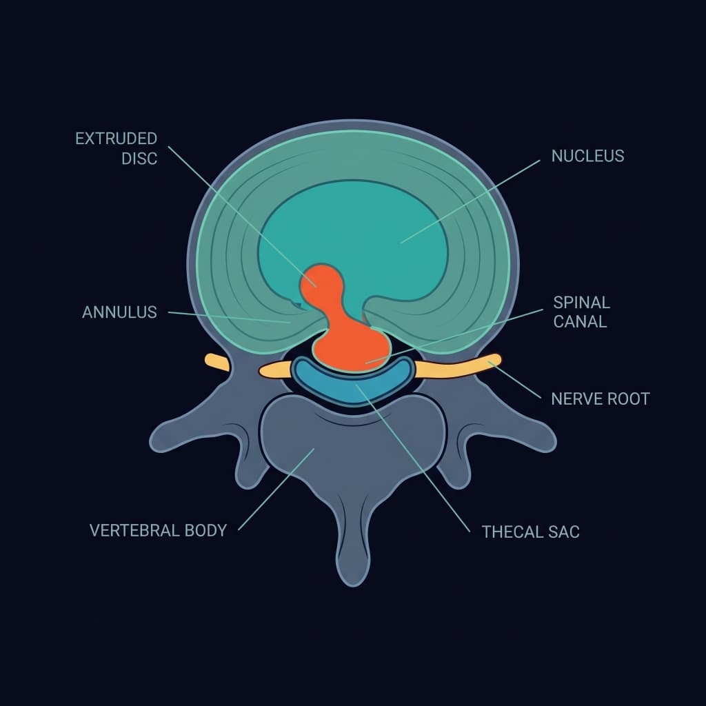
Extruded Herniated Disc
Sciatica & Radiculopathy Pain Radiation Map
Select your compressed disc level to view the corresponding leg nerve distribution path
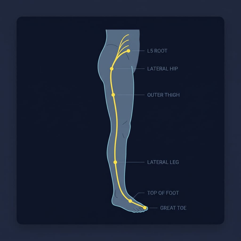
Pinched Nerve Pain Distribution & Motor Signs
Lateral Hip → Outer Thigh → Top of Foot → Great Toe
Foot Drop / Extensor hallucis weakness (difficulty walking on heels)
Tibialis posterior reflex variation
Direct 7mm decompression of the L5 Nerve Root root.
How It Works: Step-by-Step SequenceA Technical Overview of the Endoscopic Approach
A structured, ultra-minimally invasive surgical workflow designed to maximize nerve decompression while preserving normal spinal anatomy.
Micro-Portal Access
Under precise intraoperative fluoroscopy guidance, a tiny 7–10 millimeter skin incision is created.
Tubular Muscle Dilation
Sequential dilators gently separate muscle fibers along natural planes rather than cutting muscle tissue.
Endoscope Insertion
The high-definition spinal endoscope is docked at the exact anatomical site of pathology.
Direct HD Visualization
Continuous sterile saline irrigation maintains a clear, magnified field, protecting delicate neural structures.
Targeted Decompression
Micro-instruments selectively extract herniated fragments or bone spurs compressing the nerve root.
Precision Closure
The instrument is withdrawn and the tiny incision is closed with a single cosmetic suture or sterile strip.
Traditional Open Surgery vs. Endoscopic Approach
A side-by-side technical comparison illustrating the surgical differences between open and minimally invasive endoscopic techniques.
| Surgical Aspect | Traditional Open Surgery | Full-Endoscopic Spine Surgery |
|---|---|---|
| Typical Skin Incision | 50 mm – 120 mm (2 to 5+ inches) | 7 mm – 10 mm (Sub-centimeter keyhole) |
| Muscle Trauma & Dissection | Muscles stripped and detached from spinal bone | Muscles gently dilated along natural fibers without cutting |
| Bone & Ligament Removal | Extensive laminectomy often required for visualization | Preserves facet joints, lamina, and midline ligaments |
| Visualization System | Line-of-sight surgical loupes or standard overhead light | Continuous high-definition fluid-irrigated camera magnification |
| Need for Fusion Hardware (Screws/Rods) | Often required if open bone removal creates instability | Avoids fusion in appropriately selected non-instability cases |
| Postoperative Wound Soreness | Significant localized muscular soreness from detachment | Substantially less wound discomfort due to minimal tissue disruption |
| Typical Hospital Stay | 2 to 5 days of inpatient hospital recovery | Day-care (same day) or overnight observation in appropriate cases |
Clinical Disclosure: Different surgical approaches may be appropriate for different patients and conditions. While endoscopic surgery offers distinct advantages for focal disc herniations and stenosis, certain complex structural deformities or instability may still require open or instrumented stabilization techniques.
Who May Be Considered for Endoscopic Spine Surgery?Individualized Clinical Evaluation is Essential
Endoscopic surgery is not automatically suitable for every spinal condition. Thorough neurological examination and diagnostic MRI review determine clinical appropriateness.
Potential Candidates
Endoscopic procedures may be considered after comprehensive evaluation for patients presenting with:
- Patients with MRI-confirmed lumbar or cervical disc herniation causing sciatica or arm pain
- Focal spinal canal or lateral recess stenosis producing neurogenic claudication (walking fatigue)
- Symptoms that persist despite 6–12 weeks of structured conservative care (medications, physical therapy, injections)
- Patients seeking an alternative to spinal fusion or extensive muscle-stripping surgery
- Elderly or medically fragile patients who benefit from shorter operating times and reduced physiologic stress
When It May Not Be Appropriate
Endoscopic approaches may have limitations and other surgical or medical modalities may be advised for:
- ×Gross spinal instability or severe spondylolisthesis requiring corrective instrumented fusion
- ×Severe multi-level spinal deformities (e.g. progressive adult scoliosis or kyphosis)
- ×Active spinal infections (discitis, epidural abscess, or osteomyelitis) requiring open debridement
- ×Certain widespread destructive spinal tumors requiring extensive circumferential reconstruction
Endoscopic Spine Recovery Milestones
Click across the milestone timeline to inspect pain reduction curves and functional return targets
- Standing unassisted
- Light walking with physiotherapist
- Normal oral diet resumed
- Heavy lifting (>5kg)
- Prolonged sitting (>45 mins)
Frequently Asked Questions about Endoscopic Spine Surgery
Common clinical questions about anesthesia, safety, candidacy, and recovery expectations.
Consult with Dr. Achal Gupta for Precision Spine & Neuro Care
Whether you are seeking definitive relief from severe sciatica, slipped disc, or exploring minimally invasive endoscopic alternatives, we are here with ethical, compassionate guidance.